


Title
Risk assessment of plaque rupture and future cardiovascular events by multi-spectral photoacoustic imaging
Call
H2020-ICT-2016-1: Photonics KET 2016
Duration
November 2016 – October 2019
Type
RIA – Research and Innovation Actions
Technologies
photoacoustic imaging, ultrasound imaging, ultra-high power pulsed diode laser, high efficient diode laser drivers, diffractive optical beam forming, signal processing, clutter reduction.
CVENT
CVENT technology is developed in accordance to the medical needs and will lead in a commercial product ready for clinical practice.
The CVENT photoacoustic imaging system will provide a revolutionary diagnostic approach for screening, in-depth diagnosis and monitoring of carotid plaque vulnerability.
Breakthrough
CVENT will develop a compact, easy to use, non-invasive, point-of-care multimodality and multi-wavelength functional photoacoustic (PA) imaging system for quantitative diagnosis and monitoring of carotid plaque vulnerability. The system combines multimodality imaging with ultrasound (US) resolution and the functional imaging of multiwavelength light. The main effort is dedicated to reach a breakthrough in functional photoacoustic imaging to extract morphological features from the multi-wavelength PA/US data to receive functional information of plaques in the carotid artery.
Photoacoustic imaging
Photoacoustic imaging has the potential for functional imaging of plaque material but still has limited imaging depth.
Development
The interdisciplinary CVENT consortium will develop a portable beyond state-of-the-art photoacoustic imaging system with a hand-held PA/US probe for improved diagnosis of carotid plaque vulnerability. This system will provide a new diagnostic approach which is significantly superior to all existing imaging approaches. The CVENT consortium has a proven track record in developing cost effective components for portable photoacoustic imaging systems: In the FULLPHASE project (FP7 project 318067) we were able to reach already an good in-vivo imaging depth with a hand-held PA/US probe, which is currently evaluated in-vivo for the diagnosis of rheumatoid arthritis and skin diseases. Increasing the image depth for carotid plaque imaging is an ambitious goal that needs beyond-state-of-the-art development.
Carotid plaques
Vulnerable atherosclerotic carotid plaques, an age and lifestyle related vascular disease, remain a major cause of death for men and women at middle age and is also a hallmark of generalized CVD involving other critical vascular regions like the coronary arteries. Every patient exhibiting symptoms of partial occlusion of the carotid arteries is examined with ultrasound (US) because a matured carotid plaque is potentially vulnerable to rupture. When that happens, the content of the lesion entering the blood stream can cause stroke. In the EU this affects over 1 million people annually. According to the guidelines, the patient is operated on, if a 70-99% occlusion of the artery is found. However, only one out of nine operations is effective (Barnett 1998; Rothwell 2003). Even worse, 19 operations need to be done to prevent a single stroke. Hence, the current method to assess plaque vulnerability is insufficient.
Diagnosis and monitoring
From retrospective studies we know that the existence of a large lipid pool, a thin fibrous cap, and intra-plaque haemorrhages are the main indicators of rupture-prone atherosclerotic plaques in general (Finn 2010; Redgrave 2006). Recent ex-vivo application of multimodality photoacoustic imaging (Daoudi 2014) shows that imaging of different plaque components is possible (Arabul 2017). The revolutionary step forward in functional photoacoustic imaging (PA) to be made by the CVENT consortium is providing in-vivo functional imaging of vulnerable plaques in the carotid artery for screening, in-depth diagnosis and monitoring.
In-vivo diagnosis
CVENT is focused on CVD patients to screen, diagnose and monitor carotid plaque vulnerability. The risk of developing atherosclerosis, and plaque-induced stroke, increases with age and originate from an inactive and excessive lifestyle as found in Western society today and/or genetic factors. Improved diagnosis and risk assessment of plaques after initial detection will lead to a significant reduction in cardiovascular diseases risk, related disability and mortality. Subsequently, by stratifying patients into higher and lower risk groups, this will lead to a reduction in overtreatment, better allocation of healthcare cost, and thus contribute to the sustainability of the health care system in Europe.
Main Goal
The main goal of the CVENT consortium is the development of a portable multimodality,
multiwavelength photoacoustic imaging system with a hand-held PA/US probe for screening,
in-depth diagnosis and treatment monitoring of carotid plaque vulnerability by means of plaque: composition and structure, size and shape, as well as mechanical properties.
Photoacoustic imaging system engineering
- Multi-wavelength diode laser beam sources with ultra-high pulse power and short pulse duration.
- A highly efficient diode laser driver, allowing passive cooling of the hand-held probe.
- A multi-spectral diode-stack beam forming based on diffractive optical elements (DOE) integrated in a hand-held probe.
- A high-sensitivity ultrasound transducer integrated in a multi-wavelength hand-held PA/US probe.
- The CVENT ultrasound platform with a multimodality (hybrid) front-end - for US and PA data beam forming - connected to an embedded low-power-consumption high-performance computing unit based on state-of-the-art system-on-chip (SoC) technology allowing for real-time signal processing of the PA/US data.
Advanced photoacoustic imaging signal processing
- Characterize and classify plaque by multi-wavelength photoacoustic imaging and estimation of tissue composition together with the quantitative parameters speed-of-sound by ultrasound techniques, contrast in Grueneisen coefficient by thermal provocation, and mechanical properties by ultrafast plain wave strain imaging.
- Reduce measurement noise in the multi-wavelength PA images by multiple input/multiple output multi-spectral filters with adaptively optimized combinations of laser pulses.
- Suppress clutter signals that are superimposing the weaker PA signals by exploiting simultaneous US measurements, clutter spectral behaviour, and Doppler shifts of clutter from the flowing blood of large vessels.
- Improve photoacoustic imaging image quality by optimizing PA/US hybrid reconstruction techniques.
photoacoustic imaging system verification and validation
- Verify the CVENT photoacoustic imaging system in-vitro and ex-vivo using human carotid plaques obtained from surgical interventions.
- Perform a first clinical validation by imaging plaque prior to and during surgery.
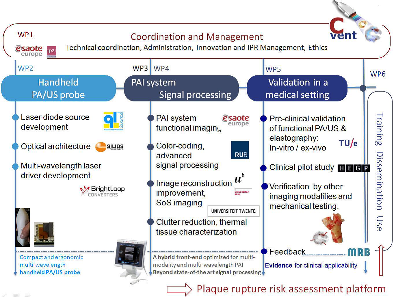
CVENT project structure
CVENT strategy
- Close involvement of physicians, from requirement specifications to the validation of the CVENT photoacoustic imaging system, with a hand-held PA/US probe.
- Short feedback loops between: medical practice, engineering and advanced signal processing during the R&D process.
- R&D on a highly sensitive PA/US probe and signal analysis/enhancement methods that warrant sufficient penetration depth and signal-to-noise ratio for in-vivo plaque imaging in the carotid artery.
- Validation of the CVENT photoacoustic imaging system in experimental and clinical settings to support an efficient translation, from research towards product development and clinical application.
- Active communication with stakeholders and patient groups to create public awareness for the CVENT approach.
In-vivo diagnosis
CVENT is focused on CVD patients to screen, diagnose and monitor carotid plaque vulnerability. The risk of developing atherosclerosis, and plaque-induced stroke, increases with age and originate from an inactive and excessive lifestyle as found in Western society today and/or genetic factors. Improved diagnosis and risk assessment of plaques after initial detection will lead to a significant reduction in cardiovascular diseases risk, related disability and mortality. Subsequently, by stratifying patients into higher and lower risk groups, this will lead to a reduction in overtreatment, better allocation of healthcare cost, and thus contribute to the sustainability of the health care system in Europe.